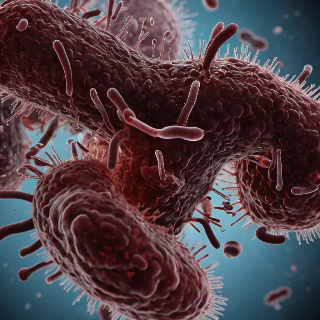
Gram staining is essential for identifying E. coli. This pink-staining, gram-negative bacterium is quickly differentiated from others, aiding in diagnosis and treatment. This article explains the gram staining process, its importance in identifying E. coli through gram staining for E coli, and tips for proper sample collection.
Key Takeaways
-
Gram staining is a critical diagnostic tool for identifying your bacteria as gram-positive or gram-negative, aiding in appropriate treatment decisions.
-
E. coli, a common gram-negative bacterium, requires prompt diagnosis through gram staining for effective management of infections like urinary tract infections and gastrointestinal illnesses.
-
Proper specimen collection and meticulous staining procedures are vital for accurate gram stain results, with common challenges including contamination and incorrect decolorization times.
Understanding Gram Staining
Gram staining is an essential diagnostic tool for identifying bacteria as gram-positive or gram-negative. This technique leverages the unique properties of bacterial cell walls to react differently to stains. Such differential staining provides microbiologists and healthcare professionals with a quick and reliable method to identify bacterial species.
In the gram staining process, bacteria undergo exposure to a sequence of stains and reagents. First, crystal violet, the primary stain, is applied, followed by iodine solution, forming a crystal violet-iodine complex within the cell. Then, a decolorizer such as ethanol or acetone is used to differentiate bacteria based on their cell wall properties. Gram-positive bacteria, with thick peptidoglycan layers, retain the primary stain and appear purple, while gram-negative bacteria lose the stain and take up a counterstain, appearing pink.
The color difference observed under the microscope categorizes bacteria into two groups with distinct characteristics and clinical implications. Gram-positive bacteria, with robust cell walls, are generally more resistant to certain antibiotics. In contrast, common gram negative bacteria, with thinner peptidoglycan layers and an outer membrane, can be more challenging to treat due to their resistance mechanisms. This distinction aids in selecting appropriate treatments and understanding the potential severity of an infection, particularly when considering gram positive organisms.
Grasping the gram reaction is crucial for accurate interpretation of gram stain results and the gram test. It offers immediate insights into bacterial identity, guiding further diagnostic tests and treatment strategies. This process exemplifies how simple techniques can yield profound results.
The Importance of Gram Staining E. coli

Escherichia coli, or E. coli, is a common gram-negative bacterium that can be both a harmless resident of the human gastrointestinal tract and a serious pathogen. Gram staining is vital for identifying E. coli, which appears pink under the microscope because it cannot retain the primary stain during the decolorization step.
Identifying E. coli through gram staining is especially important in diagnosing infections like urinary tract infections (UTIs) and gastrointestinal illnesses. As a leading cause of these conditions, rapid identification of E. coli through gram staining can greatly improve patient outcomes by guiding timely and appropriate treatment decisions. For instance, swift detection of this gram-negative bacillus in cases of coli bacteremia can be life-saving.
Gram staining is both quick and cost-effective for differentiating bacteria. Unlike advanced techniques like genome sequencing, gram stains provide immediate results, making it invaluable in clinical settings. Whether in a primary care clinic or a specialized laboratory, swiftly identifying E. coli can help curb the spread of infections, especially in cases involving contaminated food or water.
Specimen Collection for Gram Staining
Accurate gram staining results begin with proper specimen collection, where using sterile techniques to prevent contamination is paramount. Specific handling for different specimens includes:
-
Collecting sputum
-
Collecting blood
-
Collecting urine
-
Collecting throat swabs Each requires specific handling to ensure sample integrity and result accuracy.
For sample collection:
-
Sputum samples should be obtained through deep coughs into sterile containers.
-
Blood cultures are ideally drawn from two different venipuncture sites to enhance diagnostic accuracy.
-
Urine samples are best collected midstream in sterile containers to minimize contamination.
-
For throat swabs, focus on the infected area while avoiding adjacent tissues to reduce contamination risk.
Once collected, specimens must be clearly labeled and promptly transported to the laboratory to ensure viability and reliable gram stain results. Proper specimen collection, lumbar puncture, and clinical presentation is the first step toward accurate diagnosis and effective treatment.
Step-by-Step Gram Staining Procedure for E. coli

Performing a gram staining procedure involves meticulous steps to ensure accurate results, starting with slide preparation, followed by the staining process, and concluding with microscopic observation.
Each step is essential for differentiating E. coli from other bacterial species.
Preparing the Slide
Preparing the microscope slide is the first step in gram staining. Follow these steps:
-
Stir the broth culture.
-
Place 2-3 loops of E. coli onto the slide.
-
Use an inoculation loop to spread the culture into an even-thin film over a 15 mm diameter circle to ensure a uniform smear.
Allow the sample to air dry completely after spreading it. This prevents distortion of the bacterial cells during staining. Once dry, gently heat-fix the slide by passing it through a flame to ensure the cells adhere properly during staining. Incinerate any remaining bacteria on the loop after collecting the sample to maintain sterility.
Meticulously preparing the slide sets the foundation for a successful gram staining procedure. An even smear and proper heat fixation are critical for accurate and clear observation of stained bacteria under the microscope.
Staining Process
Begin the staining process with the following steps:
-
Apply the primary stain, crystal violet, to the fixed smear for one minute, staining all bacteria purple.
-
Rinse off the excess stain.
-
Apply Gram’s iodine solution to form a crystal violet-iodine complex within the cells, which helps retain the primary stain in both gram-positive and gram-negative bacteria.
Next, decolorize the slide with a mixture of ethanol and acetone to distinguish between gram-positive and gram-negative bacteria:
-
The ethanol-acetone mix dissolves the lipid layer in gram-negative bacteria, causing them to lose the primary stain.
-
Carefully monitor the duration of decolorization to avoid over-decolorizing, which can lead to false results.
Finally, apply a counterstain, commonly safranin, for one minute:
-
This stains the gram-negative bacteria pink, while gram-positive bacteria remain purple.
-
Rinse off the excess stain.
-
Allow the slide to dry before observing the results under the microscope.
Observing Results
Use oil immersion microscopy to observe the gram-stained slide. This technique enhances resolution and allows for clear visualization of the bacteria. Under the microscope, E. coli will appear as pink, rod-shaped bacilli, confirming its classification as a gram-negative organism and identifying it among gram negative bacilli. Additionally, it is important to note the presence of gram negative organisms in the sample.
Accurate interpretation of results is crucial for diagnosing infections and guiding treatment decisions. The appearance of pink, rod-shaped E. coli indicates its presence, helping differentiate it from other bacteria and aiding in the identification and management of infections.
Common Challenges and Solutions in Gram Staining

Though straightforward, gram staining comes with challenges. One common issue is thick smears, which can lead to inconsistent and hard-to-interpret results. Ensuring the smear is even and thin is crucial for obtaining clear and accurate observations under the microscope.
Another challenge is the duration of the decolorization step. Prolonged exposure to the decolorizer can remove stains from both gram-positive and gram-negative bacteria, leading to false results. Carefully monitoring decolorization time is crucial to maintain the integrity of the staining process.
Contamination is another significant concern. Non-sterile specimen collection and improper techniques can introduce contaminants, skewing results and leading to incorrect diagnoses. Ensuring specimens are collected using sterile techniques and handled properly throughout the process is key to preventing contamination. Additionally, excessive washing between staining steps can result in the loss of critical information on the slide.
Clinical Significance of Gram Staining E. coli
Gram staining serves as an essential preliminary test for identifying E. coli, especially in clinical settings where rapid diagnosis is crucial. The presence of E. coli in gram-stained specimens often indicates urinary tract or gastrointestinal infections, making this technique invaluable for initial diagnostics and immediate patient management.
By differentiating between various E. coli strains, gram staining can help identify pathogenic strains that may lead to severe illness, such as hemolytic uremic syndrome or infectious diarrhea and diarrheal illness. Quickly identifying these strains allows healthcare professionals to take prompt actions, potentially preventing disease progression and improving patient outcomes.
Immediate insights from gram staining can guide empirical antibiotic therapy even before culture results are available. This rapid identification is crucial in managing acute infections like urinary tract infection, where timely treatment can significantly affect prognosis. In severe infections like coli bacteremia or septic shock, gram staining can be the first step toward life-saving interventions.
Beyond its diagnostic value, gram staining E. coli is critical for disease control. Quickly identifying the causative organism helps track infection sources, implement control measures, and prevent outbreaks, particularly in developing countries where healthcare resources might be limited.
Enhancing Laboratory Accuracy with Milerd Detoxer

To enhance laboratory accuracy and reduce the risk of E. coli contamination, the Milerd Detoxer offers a state-of-the-art solution. This innovative device uses advanced ozone and ultrasound technology to eliminate up to 99% of harmful food toxins, ensuring safer food consumption. The Detoxer removes:
-
Bacteria
-
Viruses
-
Pesticides
-
Heavy metals
-
Mold spores
-
Parasite eggs providing comprehensive protection against potential contaminants.
The Milerd Detoxer operates using ultrasonic cleaning, creating microscopic bubbles that penetrate food surfaces to effectively remove bacteria and contaminants. This method ensures thorough cleaning while preserving the nutritional quality of the food, making it ideal for everyday use.
The Detoxer package includes an adapter, a USB Type-C cable, and a manual, making it user-friendly and convenient for daily use. The device’s compact design and straightforward controls further enhance its appeal, making it a practical addition to any health-conscious household.
By incorporating the Milerd Detoxer into your routine, you can significantly reduce the risk of E. coli and other bacterial infections, ensuring that the food you consume is safe and free from harmful contaminants. This proactive approach to disease control aligns with the recommendations of the World Health Organization and other health bodies, emphasizing the importance of food safety in preventing infectious diseases society.
Summary
In summary, gram staining is a fundamental technique in microbiology, crucial for differentiating gram-positive from gram-negative bacteria like E. coli. Understanding the gram staining procedure, from specimen collection to observing results, is essential for accurate diagnosis and effective treatment of infections. Despite its challenges, mastering this technique can significantly enhance diagnostic accuracy and patient outcomes.

The integration of advanced tools such as the Milerd Detoxer can further enhance laboratory accuracy and food safety, reducing the risk of bacterial contamination. By adopting these practices, healthcare professionals and individuals alike can contribute to better disease control and improved public health. Remember, a simple stain can reveal a world of information, guiding us towards healthier and safer living.


Laisser un commentaire
Ce site est protégé par hCaptcha, et la Politique de confidentialité et les Conditions de service de hCaptcha s’appliquent.